Recent developments on 320 multi-detector CT technologies have made the volumetric acquisition of 4D high resolution cardiac images in a single heart beat possible. We present a framework that uses these data to reconstruct the 4D motion of the endocardial surface of the left ventricle for a full cardiac cycle. This reconstruction framework captures the motion of the full 3D surfaces of the complex anatomical features, such as the papillary muscles and the ventricular trabeculae, for the first time, which allows us to quantitatively investigate their possible functional significance in health and disease. We also simulate blood flow using these meshes.
.Papers
Yang Yu, Shaoting Zhang, Junzhou Huang, Dimitris Metaxas and Leon Axel, "Sparse Deformable Models with Application to Cardiac Motion Analysis", In Proc. of the 23rd biennial International Conference on Information Processing in Medical Imaging, IPMI'13, Asilomar, CA, June 2013.
Lin Zhong, Shaoting Zhang, Mingchen Gao, Junzhou Huang, Zhen Qian, Dimitris Metaxas and Leon Axel, "Papillary Muscles Analysis from High Resolution CT using Spatial-Temporal Skeleton Extraction", In Proc. of the IEEE International Symposium on Biomedical Imaging, ISBI'13, San Francisco, CA, USA, April 2013.
Mingchen Gao, Junzhou Huang, Shaoting Zhang, Zhen Qian, Szilard Voros, Dimitris Metaxas, Leon Axel, "4D Cardiac Reconstruction Using High Resolution CT Images", In Proc. of the 6th International Conference on Functional Imaging and Modeling of the Heart, FIMH'11, New York, USA, May 2011. (Best Paper Award)
4D Cardiac Images
  |
Figure.
4D Reconstruction results of left ventricle. Papillary muscles
and the trabeculae are clearly captured. |
Cardiac Motion Analysis
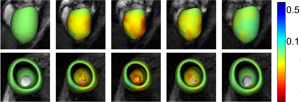 |
Figure.
The deformations of the left ventricle on a cardiac cycle
are colored by the circumferential strain |